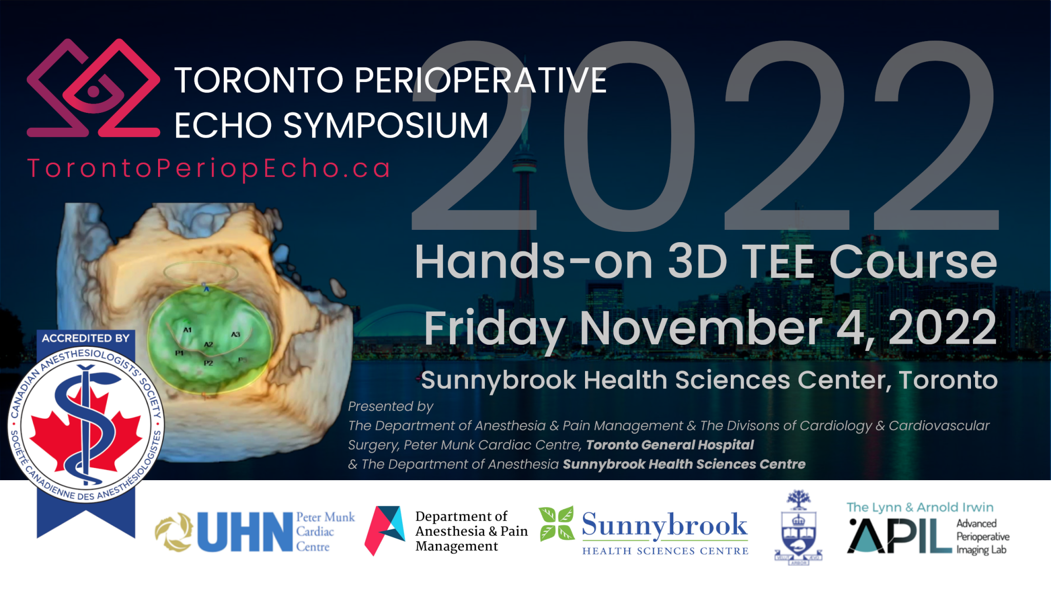
THIRD TORONTO PERIOPERATIVE 3D TEE COURSE – Hands On!
Friday, November 4th, 2022
Sunnybrook Health Science Centre, Michael Julius Atrium (M6 400)
2075 Bayview Avenue, Room M3 400, Toronto, ON, M4N 3M5
CME Accreditation
This event is an Accredited Group Learning Activity (Section 1) as defined by the Maintenance of Certification program of The Royal College of Physicians and Surgeons of Canada and approved by the Canadian Anesthesiologists’ Society. You may claim a maximum of 8.5 hours (credits are automatically calculated)
CME Planning Committee
Dr. Jacobo Moreno (Course Director), Sarah Russell, Azad Mashari, Elmari Neethling, Pablo Perez d’Empaire, Sophia Wong, Yannis Amador
Course Objectives
With improvements in the underlying technology and usability, 3D TEE is becoming increasingly integrated into the perioperative evaluation of patients undergoing cardiac and other complex surgical and minimally invasive procedures.
After active participation in this one-day course participants will be able to
- Explain the basic process of 3D TEE image acquisition and its associated limitations and pitfalls as they relate to common clinical scenarios
- Describe and demonstrate the step-by-step process of 3D TEE image acquisition and optimization for perioperative assessment of cardiac chambers and valves.
- Describe and demonstrate basic manipulation of 3D data-sets for a structures qualitative evaluation of key structures.
- Demonstrate the quantitative analysis of 3D data-sets of key cardiac structures and describe the indications and clinical relevance of 3D quantification in common clinical situations.
Program & Session Objectives
7:30 Registration & Breakfast
7:50 Introduction to Course and Faculty – Jacobo Moreno
Introduction to 3D TEE – Moderator Elmari Neethling
8:00 Anatomy for 3D echocardiography – Azad Mashari
- Describe the structure and orientation of the heart base in relation to standard TEE imaging windows
- Identify clinically relevant structures and orientation in standard 3D TEE views
- Be able to adjust probe position to optimize 3D view without going back to 2D images
8:20 Introduction to 3D TEE – Wendy Tsang
- Discuss the clinical utility of 3D Echocardiography technology.
- Explain basic principles of 3D echo imaging with modern matrix array transducers
- Describe the main 3D acquisition modes and compare and contrast their clinically relevant features and use cases (Live/Birds view vs 3D Zoom/Medium volume vs Full Volume/Large volume)
8:40 3D TEE image optimization – Jacobo Moreno
- Describe the basic function of key parameters for optimizing image acquisition: Sector Size, Depth, Gain, Focus, Line Density, Frequency, Gating, Resolution, Frame Rate.
- Describe the basic function of key parameters for optimizing 3D image rendering (post processing): brightness, chrome maps, smoothing
- Explain the diagnostic limitations, common artifacts and their mitigation strategies in 3D TEE.
9:00 Q&A
9:05 Refreshment break (10 min)
3D Acquisition: Theory & Hands-on – Moderator Pablo Perez d’Empaire
9:15 3D Acquisition: Left and Right Ventricle – Ben Walker
- Describe indications for 3D imaging of the right and left ventricles.
- Explain how to obtain 3D images of the right and left ventricles.
- Discuss the limitation of 3D vs 2D imaging of the ventricles.
9:35 3D Acquisition: Mitral Valve – Wendy Tsang
- Describe the indications for 3D imaging of the mitral valve.
- Explain the steps involved in acquiring and optimizing 3D images of the mitral valve
- Describe the qualitative and quantiative examination of 3D MV image:
- 3D views for qualitative assessment
- Cropping techniques
- Quantitative assessment
- Multi-Plain Reformating
9:55 3D Acquisition: Aortic Valve – Yannis Amador
- Describe the indications for 3D imaging of the aortic valve.
- Explain the steps involved in acquiring and optimizing 3D images of the aortic valve
- Describe the qualitative and quantiative examination of 3D AV image:
- 3D views for qualitative assessment
- Cropping techniques
- Quantitative assessment
- Multi-Plain Reformating
10:15 3D Acquisition: Tricuspid Valve – Sankalp Sehgal
- Describe the indications for 3D imaging of the tricuspid valve.
- Explain the steps involved in acquiring and optimizing 3D images of the tricuspid valve
- Describe the qualitative and quantiative examination of 3D TV image:
- 3D views for qualitative assessment
- Cropping techniques
- Quantitative assessment
- Multi-Plain Reformating
10:35 Q&A
10:40 Refreshent Break (10 min)
10:50 Hands-on acquisition
- Demonstrate the step-by-step acqusition and optimization of 3D views of left and right ventricles, mitral valve, tricuspid valve and aortic valve
12:20 Lunch Break (45 min)
- Replenish the vast amount of energy spent on the intensive learning experience of the previous 4.5 hours.
3D Clinical applications – Moderator Sophia Wong
13:05 3D TEE Clinical applications: Left and Right Ventricle– Ben Walker
- Review the importance of 3D echocardiography in the assessment of left and right ventricle during cardiac surgeries.
- Understand the importance of the use of 3D for these procedures.
13:25 3D TEE Clinical applications: Mitral Valve – Ahmad Omran
- Review the importance of 3D echocardiography in the assessment of mitral valve surgeries.
- Understand the importance of the use of 3D for these procedures.
13:45 3D TEE Clinical applications: Aortic Valve – Yannis Amador
- Review the importance of 3D echocardiography in the assessment of aortic valve surgeries.
- Understand the importance of the use of 3D for these procedures.
14:05 3D TEE Clinical applications: Tricuspid Valve – Sankalp Sehgal
- Review the importance of 3D echocardiography in the assessment of tricuspid valve surgeries.
- Understand the importance of the use of 3D for these procedures.
14:25 Q&A
14:30 Refreshment Break (10 min)
14:40 3D TEE Clinical applications: Paravalvular Leaks – Ahmad Omran
- Review the importance of 3D echocardiography in the assessment of paravalvular leaks after valve surgery.
- Understand the importance of the use of 3D for the assessment of paravalvular leaks, including localization and quantification.
15:00 3D TEE Clinical applications Mitral Clip/LAA Closure – Edgar Hockmann
- Review the importance of 3D echocardiography in the assessment of the LAA and MV during this surgery.
- Understand the importance of the use of live 3D to guide and help during this surgeries .
15:20 Q&A
15: 25 Break (10 min)
Computer analysis hands-on – Moderators Jacobo Moreno & Yannis Amador
15:35 3D image measurements and post processing analysis Lab 1: QLAB – Yannis Amador
- Review the importance of 3D images postprocessing analysis.
- Perform basic 3D measurements as areas and distances, semiquantitative calculation of 3D ejection fraction of the left ventricle, and 3D valve quantification.
16:35 Refreshment Break (10 minutes)
16:45 3D image measurements and post processing analysis Lab 2: EchoPAC – Jacobo Moreno
- Review the importance of 3D images postprocessing analysis.
- Perform basic 3D measurements as areas and distances, semiquantitative calculation of 3D ejection fraction of the left and right ventricle, and 3D valve quantification.
17:45 Summary & Concluding remarks – Jacobo Moreno
18:00 Adjournment – Jacobo Moreno
Faculty
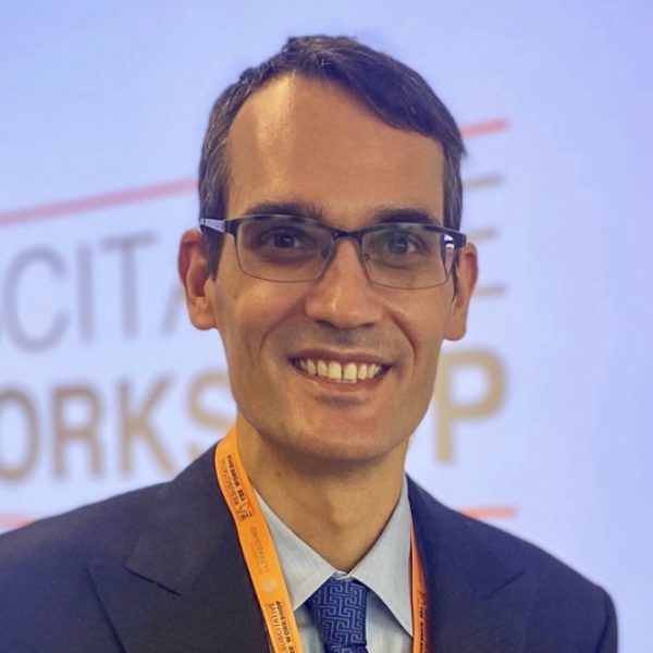
Jacobo Moreno Garijo MD MSc EDA FASE (Course Director)
Staff Cardiovascular Anesthesiologist, Department of Anesthesia, Sunnybrook Health Sciences Centre. Clinician-Investigator, Department of Anesthesia and Interdepartmental Division of Critical Care Medicine, University of Toronto. Dr. Moreno completed clinical fellowships in Critical Care Medicine (2008-2009), Cardiovascular Anesthesia (2013) and Thoracic Anesthesia (2014) at Toronto General Hospital. He is a Fellow of the American Society of Echocardiography (FASE), and is certified in Advance Perioperative Echocardiography (Advanced PTEeXAM) from the National Board of Echocardiography. He is an associate investigator at the Lynn & Arnold Irwin Advance Perioperative Imaging Lab (APIL) at Toronto General Hospital and is an active member of The Resuscitative TEE project and Perioperative Echocardiography Group at Sunnybrook Health Sciences Centre. He is the Director of the Toronto and Barcelona 3D Perioperative Echocardiography TEE courses and a long-standing member of the planning committee of the Toronto Perioperative Echo Symposium.

Edgar Hockmann MD FRCPC
Staff Cardiovascular Anesthesiologist, Department of Anesthesia, Sunnybrook Health Sciences Centre. Staff Physician, Critical Care Medicine, Scarborough Health Network. Assistant Professor, Dept of Anesthesiology & Pain Medicine, University of Toronto. Dr. Hockmann's academic focus is on the development and teaching of point of care ultrasound in the clinical setting and has been a strong advocate for providing meaningful clinical instruction to already graduated and practicing clinicians. At the University of Toronto, he has been a pioneer in the education of point of care ultrasound within critical care and anesthesia. He is certified in Advance Perioperative Echocardiography (Advanced PTEeXAM) from the National Board of Echocardiography.
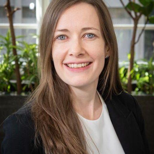
Elmari Neethling MD FRCPC
Staff Cardiovascular Anesthesiologist & Critical Care Physician, Department of Anesthesia & Pain Management, Toronto General Hospital, University Health Network. Assistant Professor, Dept of Anesthesiology & Pain Medicine, University of Toronto. Dr. Neethling graduated from Stellenbosch University and completed residency training at the University of Cape Town, South Africa. She subsequently completed both Cardiac Anesthesia and Critical Care Medicine Fellowships at the Toronto General Hospital in Canada and is certified in Advance Perioperative Echocardiography (Advanced PTEeXAM) from the National Board of Echocardiography. Her clinical and research interests in trans esophageal echocardiography, critical care medicine, perioperative outcome and point-of-care ultrasound (POCUS).
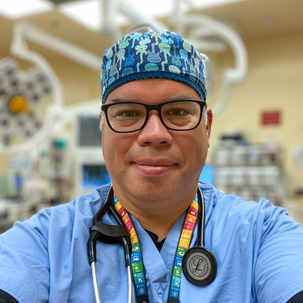
Pablo Perez D’Empaire MD FRCPC FCCM FASE
Staff Cardiovascular Anesthesiologist, Department of Anesthesia, Sunnybrook Health Sciences Centre. Lecturer, Dept of Anesthesiology & Pain Medicine, University of Toronto. Dr. Perex D'Empaire completed his undergraduate medical educaition at the University of Zulia, Venezuela. He completed his residency in anesthesiology and clinical fellowships in Critical Care Medicine and Cardiovascular Anesthesiology at the University of Toronto. He is a Fellow of the American College of Critical Care Medicine (FCCM) and the American Society of Echocardiography (FASE). He is certified in Advance Perioperative Echocardiography (Advanced PTEeXAM) and Critical Care Echocardiography (CCEeXAM) by the National Board of Echocardiography.

Wendy Tsang MD FRCPC FASE
Staff Cardiologist and Clinician Investigator, Toronto General Hospital, University Health Network. Associate Professor, Department of Medicine, University of Toronto. Dr. Tsang is the head of the Complex Valve clinic at Toronto General Hospital. She completed her medical degree at Queen’s University and went on to complete her Adult Cardiology fellowship at the University of Toronto. She then completed a CIHR-funded Multimodality Cardiac Imaging research fellowship at the University of Chicago. During that time, she also obtained a Master’s degree in Health Studies. She was a member of the writing groups for both the 2012 ASE/EACVI 3D Guidelines and the 2015 ASE/EACVI Chamber Quantification Guidelines. She also serves on the Editorial Board of the Journal of the American Society of Echocardiography and is a member of the Royal College of Physician and Surgeon’s Area of Focused Competency subcommittee in Adult Echocardiography. She currently holds a Heart and Stroke Foundation of Canada National New Investigator Award. Her research focuses on congenital and valvular heart disease, 3D echocardiography, and artificial intelligence.
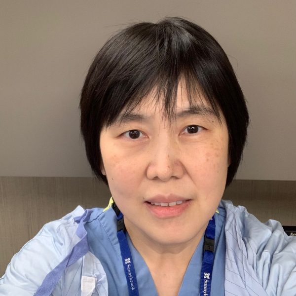
Sophia Wong MD FRCPC
Staff Cardiovascular Anesthesiologist, Department of Anesthesia, Sunnybrook Health Sciences Centre. Associate Professor, Dept of Anesthesiology & Pain Medicine, University of Toronto. Dr. Wong is the director of Cardiac Anesthesiology at Sunnybrook Sciences Health Centre with over 20 years experience in perioperative echo.
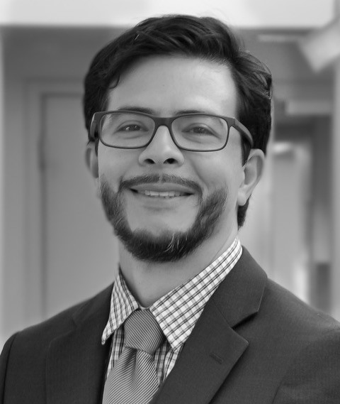
Yannis Amador MD FRCPC
Staff Cardiovascular Anesthesiologist, Department of Anesthesiology & Perioperative Medicine, Kingston Health Sciences Centre Assistant Professor, Department of Anesthesiology, Queen's University. Dr. Amador completed residency training in anesthesiology and emergency medicine in Costa Rica. Subsequently he completed a research fellowship with Dr. Feroze Mahmood at Beth Israel Deaconess Medical Center, Harvard Medical School followed by a clinical fellowship in cardiovascular anesthesiology at Toronto General Hospital. He is certified in Advance Perioperative Echocardiography (Advanced PTEeXAM) from the National Board of Echocardiography. He is the organizer of the ulti-disciplinary echo rounds at the Kingston Health Sciences Centre and a member of the organizing committee for the POCUS workshop at the Canadian Anesthesia Society (CAS) meeting.
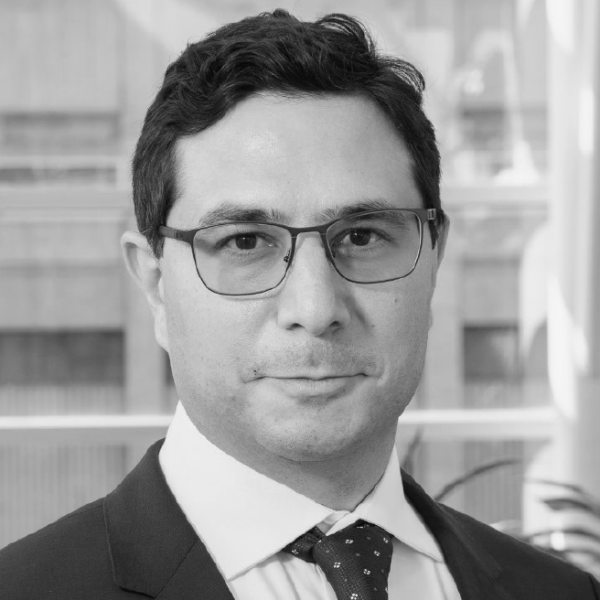
Azad Mashari MD FRCPC
Staff Cardiovascular Anesthesiologist, Department of Anesthesia & Pain Management, Toronto General Hospital, University Health Network. Assistant Professor, Dept of Anesthesiology & Pain Medicine, University of Toronto. Dr. Mashari completed his residency and fellowship training in cardiovascular anesthesiology at the University of Toronto. He is the director of the Advanced Perioperative Imaging Lab (APIL) at Toronto General Hospital and chair of the planning committee for the Toronto Perioperative Echo Symposium. He is certified in Advance Perioperative Echocardiography (Advanced PTEeXAM) from the National Board of Echocardiography. His research focuses on applications of medical image computing, 3D modeling and 3D printing in medical education and perioperative care.
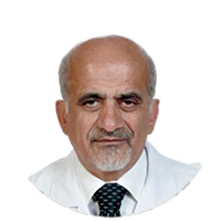
Ahmad Omran MD, FACC, FESC, FASE, FECVI
Staff Cardiologist, Toronto General Hospital, University Health Network. Dr. Omran completed his fellowship in echocardiography at Toronto General Hospital in 1996 and subsequently collaborated with Tirone David in developing intraopertive imaging techniques to support the development of novel techniquest for aortic and mitral valve repair. He is the author of landmark papers on TEE assessment of mitral valve anatomy and suitability for mitral valve repair and created the first map of TEE classification of the mitral valve segments and scallops which is still in use in many centers. Dr. Omran has published more than 35 papers in role of TEE in valves repair and contributed chapters in 5 textbooks of echocardiography including chapter of role of 3D TEE in cardiac operating room in the “Nanda’s Comprehensive Textbook of Echocardiography”. He is certified by National Board of Echocardiography and European Association of Cardiovascular Imaging (EACVI) and is an active member of American and European societies of echocardiography. He is currently working as staff cardiologist supporting the perioperative echocardiography program at Toronto General Hospital. He is a highly regarded teacher in echocardiography and runs the quality assurance program for intraoperative TEE at Toronto General Hospital.
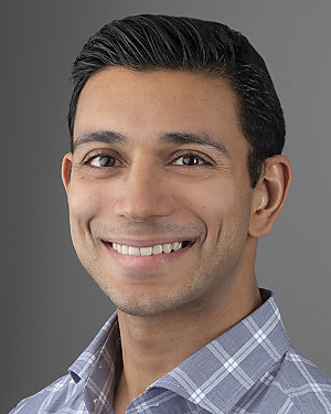
Sankalp Sehgal MD
Staff Cardiac Anesthesiologist, Department of Anesthesiology, Critical Care and Pain Medicine, Beth Israel Deaconess Medical Center (BIDMC), Harvard Medical School, Boston Dr. Sehgal specializes in anesthesia for cardiothoracic surgery, vascular and thoracic procedures, and is a member of the structural heart program. His research focuses on cardiothoracic surgical outcomes, advanced perioperative echocardiography as well as cardiac electrophysiology lab. He is a a co-investigator the TRILUMIATE and TRISCEND trials for tricuspid valve interventions. He is the former associate vice-chair of education (2014-16), section head for peri-operative echocardiography (2014-18) and mentor for the Annual New York TEE conference sessions (2016-18). He has been an invited panel speaker at numerous annual national and international conferences including the Indian Association of Cardiovascular and Thoracic Anesthesiologists (IACTA) International TEE Webinars, the American Society of Anesthesiologists (ASA) conference, New York State Society of Anesthesiologists (NYSSA) PGA conference and the Annual Perioperative and Critical Care Echocardiography workshop, India.
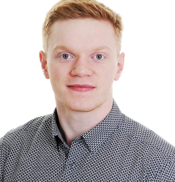
Ben Walker MD
Dr. Walker was born and raised in Ottawa, Ontario, where he spent most of his youth mountain biking and skiing in the Gatineau Parc hills. In the pursuit of outstanding medical training, skiing, and mountain biking, he decided to train in Utah for both his Anesthesiology Residency and Cardiothoracic Fellowship.
Competing Interests Policy
Faculty will declare all relevant relationships and potential competing interests during their presentation.
Faculty COI statements will be posted here when available.
Should a conflict of interest be identified a variety of strategies will be employed for mitigation, as recommended by the Royal College of Physicians and Surgeons of Canada (https://www.royalcollege.ca/rcsite/cpd/accreditation/toolkit/faqs-on-accreditation-e ). These include revision of the presentation topic or focus; peer review of the content in question to ensure scientific integrity and balance. If the concern raised is not addressed by these methods, an alternate speaker for the topic will be sought out, or the topic removed from the program.
Share this:
- Click to share on Telegram (Opens in new window) Telegram
- Click to share on WhatsApp (Opens in new window) WhatsApp
- Click to share on Facebook (Opens in new window) Facebook
- Click to share on X (Opens in new window) X
- Click to share on LinkedIn (Opens in new window) LinkedIn
- Click to email a link to a friend (Opens in new window) Email
